• Skip to content
  • Training Programs
    • /r/bodyweightfitness Recommended Routine (Free; Beginners)
    • Intermediate Bodyweight Training (Free; Push/Pull Split)
    • My Rings Routine (Purchase Here)
    • My Hypertrophy Routine (Purchase Here)
  • Articles & Tutorials
    • Fitness Index
    • Nutrition Index
    • Inspirational Index
    • Science Index
      • Human Anatomy Series
      • Human Physiology Series
      • Pharmacology Series
  • About Me
  • Members Area
    • Login
    • Account
    • Rings Routine (Members Only)
    • My Hypertrophy Routine (Members Only)
    • Slackline Program (Members Only)

Antranik.org

Strength & Flexibility Expert

Like! Follow! Subscribe!

  • Facebook
  • Instagram
  • YouTube

Function of the Atrioventricular and Semilunar Valves

By Antranik 2 Comments

When we have blood coming in from the atria, an atrial contraction is going to try to really squeeze all the blood, including the last bit of it so that the ventricles are very, very full so that you could be more efficient.  Over the course of a day this efficiency adds up.

When the two ventricles are full, they are going to contract and the blood is going to go the only way it can, to the vessels above.  When the blood goes up, the blood is going to try to push back down due to gravity but because the shape of the tricuspid and bicuspid valves are somewhat like umbrellas, when the blood pushes up against them, it closes them shut.  When the ventricles are done contracting, the atrioventricular valves relax and that takes the pressure away cause of the blood that was just pushed into the vessels. There is a tendency for the blood to fall back down, but because of the shape of the semilunar valve, the valves hit each other and so that’s your second sound from your heart beat.


The Fibrous Skeleton

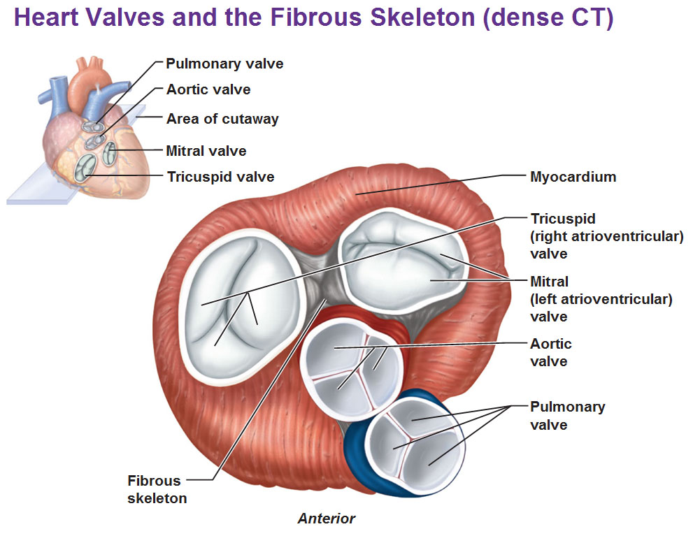
This particular view below slices the heart at the level where you could see all of those valves, from the top and looking in to get a view of the arrangement of these cusps.  Observe the tricuspid valve with its three cusps/cups.  And the right side (left anatomically) is the mitral bicuspid valve.  You see the aortic valve and pulmonary valve don’t really look like half moons, they look like pies but we can’t call them the aortic pie slice, so anyway, that’s what they are.

You could see encircling each valve, is pretty dense connective tissue and that’s on purpose because the valves have to withstand quite a bit of pressure over a course of a lifetime.  Blood is constantly swishing past by and when the ventricles contract, the valves end up getting slammed shut and so this very dense fibrous tissue has to keep the shape of the valves and anchor the cusps.  It also prevents overdilation meaning it prevents them from getting too stretched out.  Also another concept is that the fibrous tissue helps block the spread of electrical impulses.  We’re going to have impulses running all over the heart but we want the direction of the impulses to be very specific so everything moves in the right direction.  So at certain points the spread of electricity gets blocked by this fibrous skeleton so the electricity is not going every which way.

Use this Table of Contents to go to the next article

Painting by Michael Reedy
The Basics. Start here. This is your Foundation.
  • Intro to human anatomy
  • The building blocks of cells

Epithelial and Connective Tissue

  • Basics of Epithelial Tissue
  • Eight types of Epithelial Tissue
  • Detailed Features of Epithelia
  • Connective Tissue Basics
  • Three Types of Membrane

Skin, Hair, Nails, Sweat Glands

  • Integumentary System Part 1
  • Integumentary System Part 2

The Skeletal System

  • Cartilage and Bones
  • Geography of the Skull
  • Special parts of the skull
  • The Vertebral Column
  • Thoracic Cage, Ribs, Fontanelles

The Muscular System

  • Muscles of the Head
  • Muscles of the Neck and Vertebral Column
  • Muscles of the thorax for breathing and the pelvic floor (The Diaphragm)
  • Muscles of the Abdominal Wall
  • Muscles of the Forearm

The Central Nervous System

  • Fundamentals of the Nervous System and Nervous Tissue
  • CNS: Intro to Brain and Ventricles, Medulla Oblongata, Pons, Mid-Brain and Cerebellum
  • Central Nervous System: Spinal Cord
  • The Diencephalon
  • The Cerebral Hemispheres
  • Functional Areas of The Cerebral Cortex
  • Cerebral White Matter and Gray Matter and Basal Ganglia
  • The Limbic System and the Reticular Formation
  • Protection for the Brain: Meninges, CSF, Blood-Brain Barrier
  • Disorders of the Central Nervous System

The Peripheral Nervous System

  • Peripheral Nervous System: Cranial Nerves
  • Peripheral Nervous System: Spinal Nerves and Plexuses
  • Innervation of the skin: Dermatomes

The Autonomic Nervous System

  • The Autonomic Nervous System (Includes sympathetic and parasymphathetic systems)
  • Visceral Sensory Neurons and Referred Pain

Special Senses

  • Chemical Sense: Taste (Gustation)
  • Chemical Sense: Smell (Olfaction)
  • The Eye and Vision

YOU ARE HERE AT THE CARDIOVASCULAR SYSTEM

The Cardiovascular System

  • Intro to the Heart
  • Blood Flow of the Heart (Circulation Flow)
  • Myocardium
  • Conducting System of the Heart (Nervous Impulse Flow)
  • The Four Corners of the Heart
  • Layers of the Pericardium, Heart Wall and Spiral Arrangement
  • Function of the Atrioventricular and Semilunar Valves
  • Blood Components, Hemoglobin, Type/Rh Factor, Agglutination
  • Blood Vessels

Specialized Systems

  • The Endocrine System (Pituitary, Thyroid, Pancreas, Adrenal, Gonads, etc)
  • The Lymphatic System (Spleen, Thymus, Lymphatic vessels, nodes etc)
  • The Respiratory System (Lungs, Alveoli, Bronchi, Trachea, Larynx, Nasal cavities, etc)
  • The Immune System
  • The Urinary System: Kidneys
  • The Urinary System: Ureter and Urinary Bladder


Other posts you may enjoy:

  1. Cartilage and Bones
  2. Thoracic Cage, Ribs, Fontanelles
  3. The Diencephalon
  4. Intro to the Heart
  5. The Lymphatic System

Filed Under: Anatomy, Science

Contact · Log in