We have 3 layers of muscles that prevent the organs from falling down. Colloquially we call this the diaphragm! Note that the “perineum” is the diamond-shaped area defined by these four landmarks: Public symphysis, coccyx, ischial tuberosities. The perineum includes all structures inferior to, or external to the pubic diaphragm. Not to be confused with peritoneum which is the peritoneal cavity.
Superior Pelvic Diaphragm
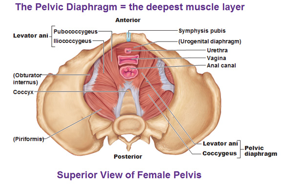
The superior pelvic diaphragm is the inner, deepest layer.
The superior pelvic diaphragm is made of two layers called the Coccygeus and the Levator Ani which create the pelvic floor.
Coccygeus originates from the ischial spine and inserts into the coccyx (and lower sacral margin.)
The Levator Ani layer is actually a pair of muscles called…
- pubococcygeus (pubis to coccyx)
- iliococcygeus (very bottom of iliac bone to coccyx).
These muscles flatten out when it contracts to allow the lungs to expand and take in air.
The urogenital diaphragm
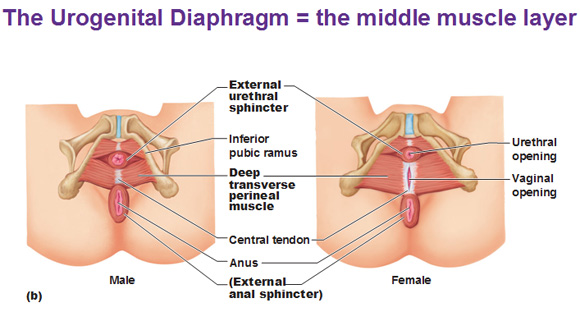
This is the second layer that is made of 2 muscles called the external urethral sphincter and deep transverse perineal muscle.
External urethral sphincter: This surrounds the urethra and voluntarily constricts to prevent you from peeing. Since it is a sphincter muscle, it is round, but not only is it round, it is a skeletal muscle, which means we have voluntary control over this.
Deep transverse perineal: This runs from the ischial rami to the central tendon of the perineum, which is located at the exact center of the perineum. This helps support the pelvic organs.
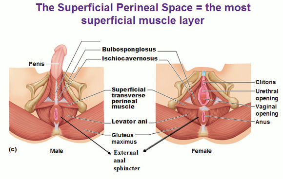
Muscles of the superficial perineal space
The superficial layer is the layer most inferior when the pelvis is upright (as opposed to the superior pelvic diaphragm). It contains 4 muscles:
the superficial transverse perineal muscle (most bottom) strengthens the central tendon and runs from ischial tuberosities to central tendon.
The ischiocavernosus (runs along side ischium) and the bulbospongiosus (kind of surrounding/supporting the base of penis or either side of the vagina) maintains erections for the penis or clit.
The external anal sphincter (again, a skeletal muscle, which is voluntary, and that’s why we have to learn to poop) prevents defecation.
 Deep Muscles of the Thorax for Breathing
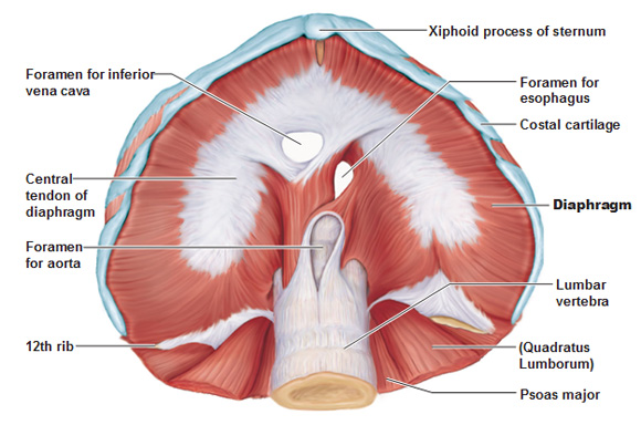
The diaphragm we just discussed: In regards to the picture below.. If you tilt a skeleton and look UP at the rib cage from the bottom, that’s what you’re seeing. The ribs and xiphoid process are on the top. The diaphragm is shaped like a dome at rest. When this muscle contracts we create more space/volume because the muscle goes down and flattens and takes air in. When you exhale the diaphragm relaxes.
We definitely need blood and food to bypass this part of the body, that’s why we have a couple holes in this muscle to let structures run through. The foramen (hole) inferior vena cava drains blood from the legs to the heart. The foramen of the esophagus allows food to pass down to the stomach. The foramen for the aorta allows the blood to enter the body from the heart.
External and Internal Intercostals
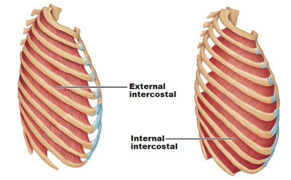
The external intercostals origin is the inferior border of the rib above.
The insertion is the superior border of the rib below.
The fibers run diagonally downward from lateral to medial. (just like the external obliques, just think of how you put your hands in your pocket, that’s the direction the fibers run for the externals)
Function? They pull the ribs up to elevate the rib cage. When these contract you get chest breathing. This kicks in when you go running, even though, ironically most people do shallow breathing with the chest at rest, rather than belly breathing.
The internal intercostal origin is the superior border of the rib below.
The insertion is the inferior border of the rib above.
The fibers run diagonally downward from medial to lateral. (just like the internal obliques, opposite of the external obliques)
Function? You guessed it, pulls the ribs down to depress the rib cage.
Use this Table of Contents to go to the next article
YOU ARE HERE AT THE MUSCULAR SYSTEM
