The Urinary System: Ureter and Urinary Bladder
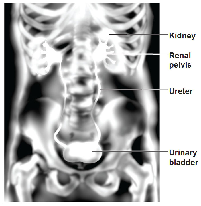
Ureters are 10 inch long tubes that carry urine from kidneys into urinary bladder. Their oblique entry into the bladder prevents backflow of urine and increased pressure within the bladder compresses the distal ends of the ureters shut.
 This picture is a pyelogram that shows the urinary bladder. They’ve injected a dye into the system and give it time to work its way to the kidneys and when it is splatted through the glomeruli and not taken back into the blood stream through the peritubular capillaries or vasa recta, it goes to the calices and the renal pelvis and from there drains into the ureters. Then they take a special x-ray that shows that dye.
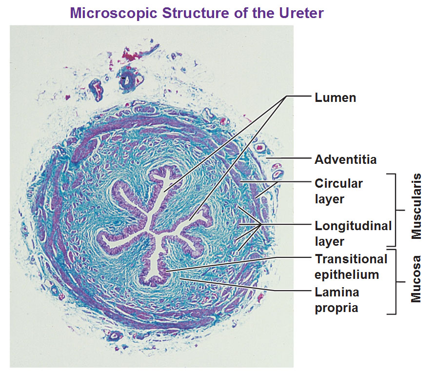 Above: Here’s a cross section of a ureter. See it’s not a simple structure. The lumen is not even a circle, it has a bunch of folds. It’s always about surface area but in this case the folds allow urine to pass through.. When you have a bolus of liquid passing through, if you didn’t have those folds, it would rip the structure. Our mucosa is lining the lumen. The lamina propia is the basic supportive tissue and transitional epithelia? Because of its shape and the way the cells are stacked up on each other, it stretches. We know this really well because if you’re stuck in the freeway and you have to pee, but you can’t, this nice epithelium allows the inside of the bladder and ureters to do that extra stretching.
On the outside of that is a smooth muscle layer called the muscularis made up of the longitudinal layer and the circular layer running in right angle directions: The circular layer runs around the lumen while the longitudinal layer runs along the length of the ureter. This muscularis layer squeezes the urine along via peristalsis. It’s not a passive system and doesn’t depend on gravity. Then our most outer surface is a faint layer called adventia.
Urinary Bladder
The urinary bladder is a sac that stores urine and squeezes it out. When it’s full it’s actually very round and expands into the abdominal cavity. When it’s empty you can’t feel it as it lies within the pelvis.
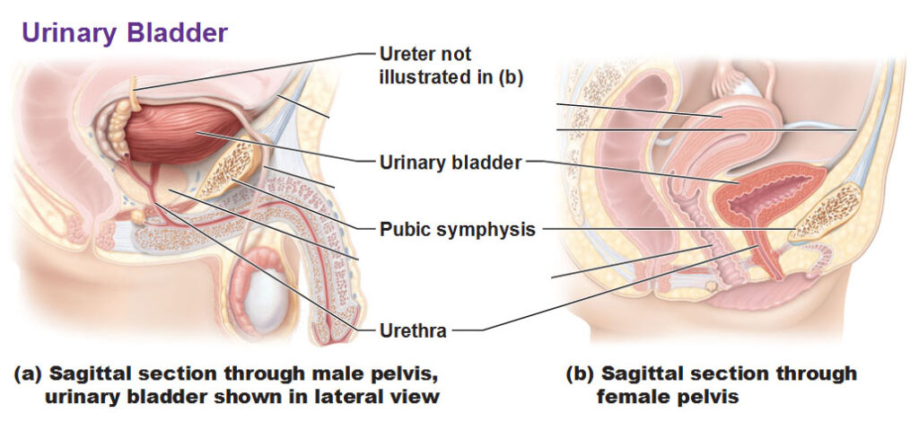
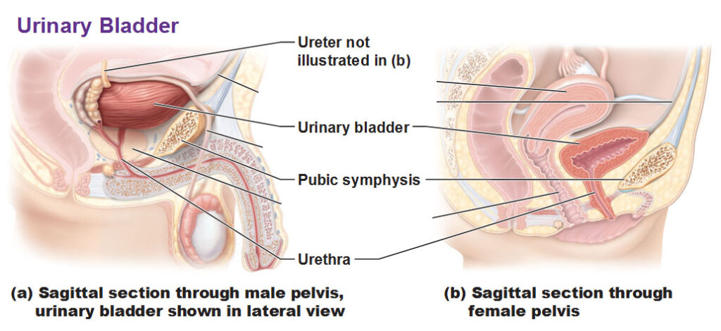 Here’s our bladder in both males and females. In the male picture you are seeing the outside surface but in the female slide you’re seeing the inside surface. You see it’s a very thick layer as it’s quite muscular. The ureter can be seen entering in at an oblique angle and eventually the urine enters the urethra.
Histology of the urinary bladder
Here’s just another slice of the bladder, somewhat similar to the layers of the ureter.
The thick layer of smooth muscle is called the detrusor muscle with the adventitia on the outside which is just dense CT.
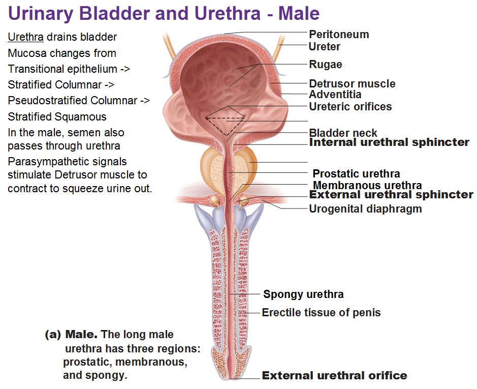
Here’s the male view:
Above: You could see the detrusor muscle making up the wall of the bladder. The rugae are the folds of bladder mucosa inside the urinary bladder that allow it to expand much much greater than its original size. Then we have the neck of the bladder. Then we have the involuntary smooth muscle called the internal urethral sphincter muscle. The external urethral sphincter muscle is voluntary skeletal muscle and the one we train from childhood to hold our pee. The prostatic urethra is the part of the urethra in the middle of the prostate. Then part of the urethra in the penis is called the spongy urethra. In between the spongy and prostatic is the membranous urethra.
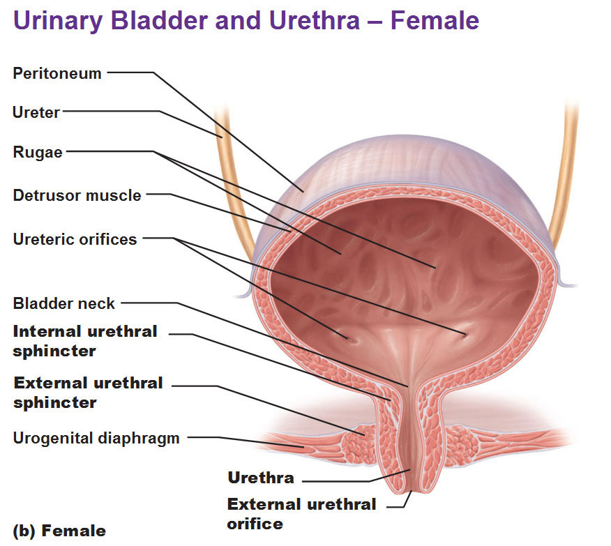
Female view:
Note that urethra is separate from vagina. The internal sphincter goes straight to the external urethral sphincter with no prostate in between and this is much shorter because there’s no penis to continue.
Use this Table of Contents to go to the next article
YOU ARE HERE AT THE SPECIALIZED SYSTEMS


