The Autonomic Nervous System
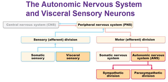
Up until now we’ve covered everything in this organizational chart below except for the ANS and visceral sensory system, which are highlighted in yellow.
 The autonomic nervous system (ANS) is a system of motor neurons that innervate smooth muscle, cardiac muscle and glands.
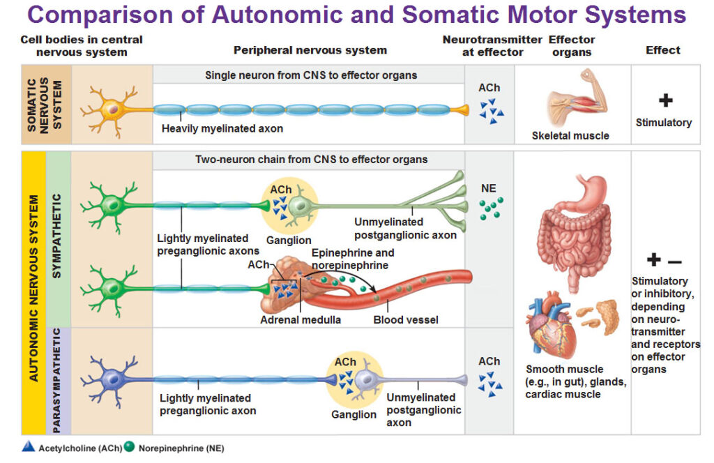
The autonomic nervous system has two divisions: Sympathetic and Parasympathetic. They mostly innervate the same structures but cause opposite effects. The sympathetic division mobilizes the body during extreme situations such as exercise, excitement and emergencies. Colloquially known as “fight or flight.” The parasympathetic division controls routine maintenance functions such as to conserve body energy and is colloquially called as “rest and digest.”
To make sense of the picture above, note the following…
A neuron found in the parasympathetic nervous system has:
LONG myelinated axon –> ganglion –> SHORT unmyelinated axon.
A neuron found in the sympathetic nervous system has:
SHORT myelinated axon –> ganglion –> LONG unmyelinated axon.
Now is a good time to make sure we differentiate that the ganglion we are talking about is not the same as the dorsal root ganglia. The “ganglia” in the ANS are motor ganglia made up of cell bodies of motor neurons. The “dorsal root ganglia” is in the sensory somatic part of the peripheral nervous system and are sensory ganglia made up of cell bodies of sensory neurons. Review the location of the dorsal root ganglia here: CNS: Spinal Cord
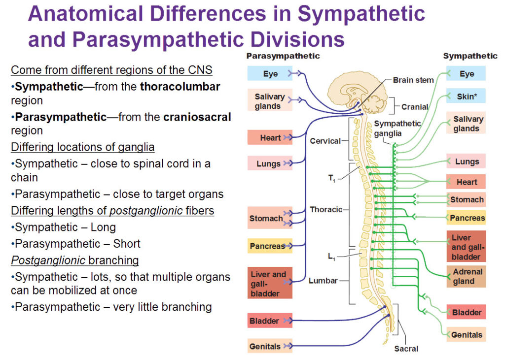
The Parasympathetic Division
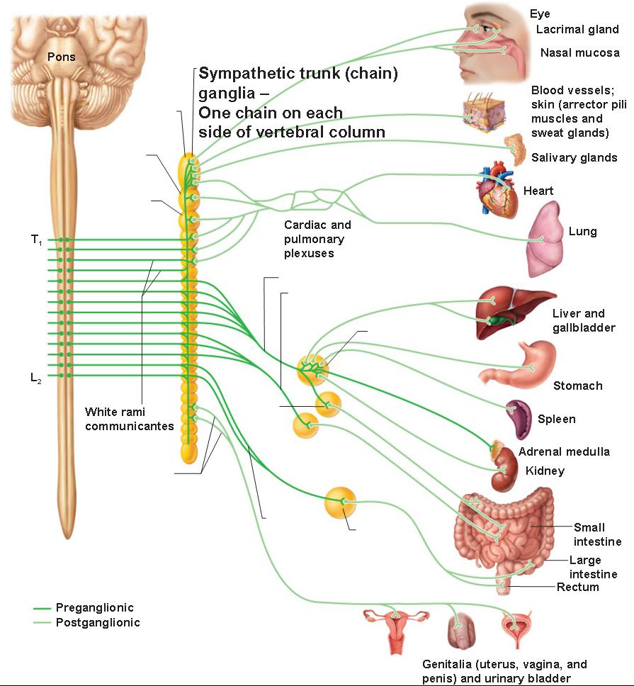
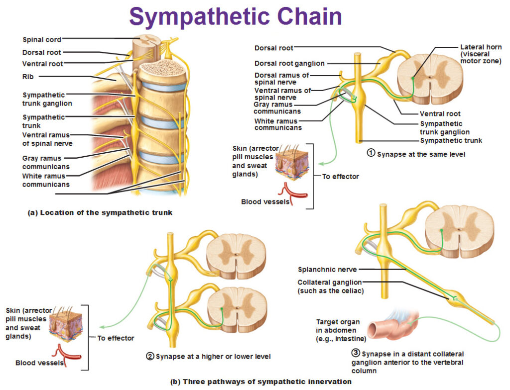
The Sympathetic Division (T1 – L2)
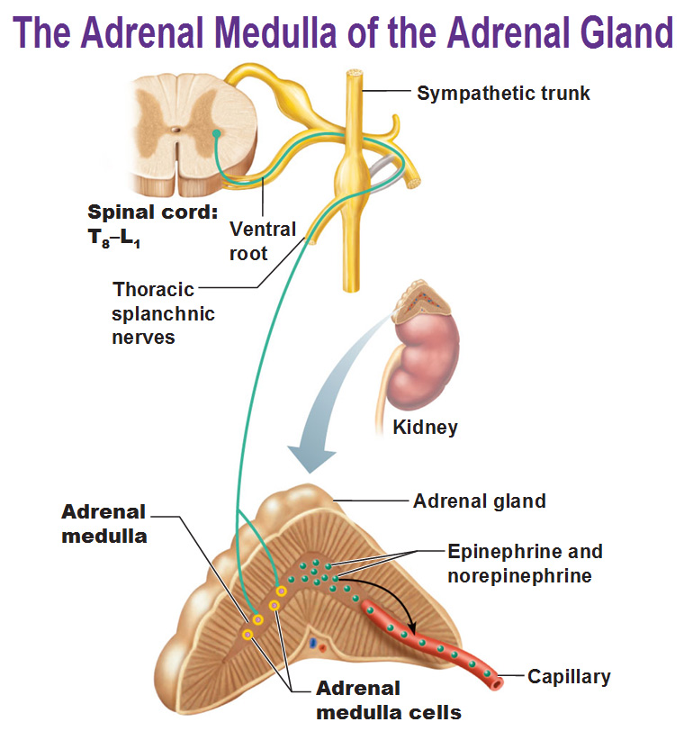
Adrenal Medulla
The adrenal medulla is GIGANTIC. It’s the largest sympathetic ganglia. The cells are made of modified neurons that have short axons and no nerve processes. The adrenal cortex is an endocrine organ and the outer layer is the adrenal cortex while the inner layer is the medulla. When stimulated by preganglionic sympathetic fibers from T8-L1, they secrete large quantities of the excitatory hormones norepinephrine and epinephrine (adrenaline) into nearby capillaries. When these two hormones are released in the blood they amplify all of this fight or flight stuff to give you more energy.
Use this Table of Contents to go to the next article

YOU ARE HERE AT THE ANS


