Integumentary System Part 1
Integumentary = Relating to Integuments. From the Latin word “integumentum” which means a covering.
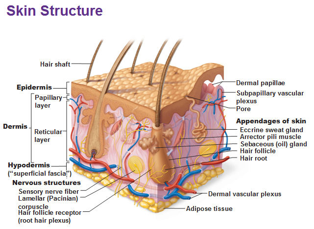
Skin is made of 2 layers: epidermis (which is epithelium tissue) and dermis. The dermis is your underlying connective tissue. The hypodermis is under the dermis that consists of adipose and loose areolar connective tissue.

Functions of Skin
1) Protection is the most important, including protection from water loss.
2) Regulates body temp via capillaries and sweat glands.
3) Excretion of urea (broken down amino acids) through sweat
4) Vitamin D
5) Sensory reception – nerve endings detect pressure, temp and pain
Keratinocytes are a type of cell (-cyte) that is most abundant in the skin. The deepest layer has lots of mitosis going on with cells producing keratin (a tough, fibrous protein). Keratin are made of microfilaments. Keratin helps keep cells together across a bunch of cells. Keratinocytes live 35-45 days and are dead by the time they are near the surface of the skin.
Epidermis
Epidermis is our epithelium layer made of stratified (a lot) squamous (squashed) epithelium. We have 4 chunks of layers called strata. We have 5 total in the palms and feet.
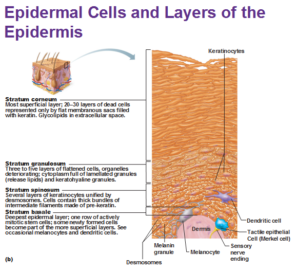
1. Stratum basale (base) = deepest SINGLE layer.
- Other than keratinocytes we have merkel cells that are always associated with a nerve cell that help you feel pressure.
- We also have melanocytes (melanin-cyte) that secrete melanin that are taken up by keratinocytes to protect themselves, like an umbrella that shields the nucleus. If the UV light gets to the nucleus it can mutate due to DNA damage.
- In lighter-skinned people, melanin is broken down by lysosomes in the more superficial cells; in darker-skinned people, melanin remains in all epidermal layers. Is this why black people are more susceptible to skin cancer from UV radiation?
2. Stratum spinosum = “spiny” appearance due to artifacts from crude microscopes.
- Keratinocytes create pre-keratin here.
- Pre-keratin is made of intermediate filaments.
- Certain dendritic cells devour (phagocytosis) foreign objects.
3. Stratum granulosom = granules.
- Keratinocytes are getting squashed at this point. The keratinocyte granules help edit pre-keratin into keratin.
- Lamellated granules are secreted into the matrix that are a waterproofing glycolipid (the witch from wizard of oz didn’t have these).
- The plasma membranes of keratinocytes start thickening here as well.
4. Stratum lucidum = under a microscope it looked thin (lucid) but this is the thick skin of palms and soles. Once you get to this distance it is too far for O2 and nutrients to get to.
5. Stratum Corneum (corneum means horn: hard like a horn): 20-30 dead layers of keratinocytes w/ thick plasma membranes are here that continually slough off. Keratin = prekeratin + glue from the granules
Before we move onto the Dermis… let’s talk about the what is a callus and why does it form?
A callus is a localized hyperplasia (overdevelopment) of the stratum corneum of the palms or soles due to pressure on the skin or friction and the consequent increase in mitotic activity of the stratum basale in that localized area. The callus is the response to this increased need for protection against mechanical abrasion.
Dermis
Papillary layer = superficial 20% = Areolar connective tissue
Dermal papillae are like little fingers that project into the epidermis to increase surface area (like microvilli).
Dermal ridges are found in feet and palms (aka friction ridges).
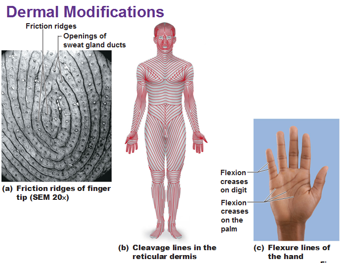
Reticular layer = Deep 80% = dense irregular connective tissue
Named for networks of collagen fibers giving its strength (not reticular fibers). Collagen is everywhere irregularly for freedom of movement.
Stretch marks are torn dermal collagen.
Flexure lines usually go over joints that show as a crease in the skin.
Cleavage lines are topographical areas on the body where the collagen tends to be thinner which is important for surgeons to know so they make incisions parallel to those lines to improve healing.
Hypodermis (aka superficial fascia aka subcutaneous layer)
This part helps anchor the skin to the underlying structures but it’s loose enough to allow skin to slide freely over. Mostly fat and areolar connective tissue and contains tons of things.
Melanin is the most common pigment. Carotene (yellowish) exists in the fat of the hypodermis (and the stratum corneum of the epidermis). Hemoglobin which gives blood its color can be seen through light skinned people.
Vitamin D is necessary for the absorption of calcium in the digestive tract.
Skin cancer (carcinoma) names depend on cell origin.
Basal cell carcinoma – From cells of stratum basale (least malignant)
Squamous cell carcinoma – From squamous cells of the stratum spinosum.
Melanoma – from melanocytes are the worse b/c melanocytes move a great distance from embryonic development b/c of their innate tendency to travel. When they are damaged, their childhood desire to be somebody and travel explodes and they go to the brain and metastasize (singing in the brain).
Use this Table of Contents to go to the next article
YOU ARE HERE


