Functional Areas of The Cerebral Cortex

We have two types of functional areas:
Sensory areas
•Primary Sensory Cortex – makes you aware of a sensation
•Association areas – give meaning to/make associations with a sensation
•Multimodal Association Areas – make associations between different types of stimuli
Motor areas – allow you to act upon a sensation
•Premotor Cortex – plans movements; then
•Primary Motor Cortex – sends signals to generate movements
•2 special motor cortices (Frontal Eye Field, Broca’s area)
Primary Sensory Cortex
For each of the major senses, there is an area called the primary sensory cortex. These are represented with dark blue in the diagrams and include somatosensory, visual, auditory, vestibular, taste, smell, visceral sensations. Something that isn’t shown is the vestibular cortex which is located in the insula, just below the temporal and frontal lobes.
Primary Olfactory Cortex
On top of the cribriform are the nasal foramina and they hit the olfactory bulb which then run toward the primary olfactory cortex through the olfactory tract. This cortex is where you get sensation of smell, before you’ve figure out what the smell is.
The olfactory cortex is located on the medial aspect of the temporal lobe, in the uncus (aka piriform lobe). The olfactory cortex is also called the Rhinencephalon, or “nose brain.” This is the most primitive part of the cerebrum and connects directly to the limbic system (emotional system), which is why smells often directly trigger emotions as well as our deepest memories.
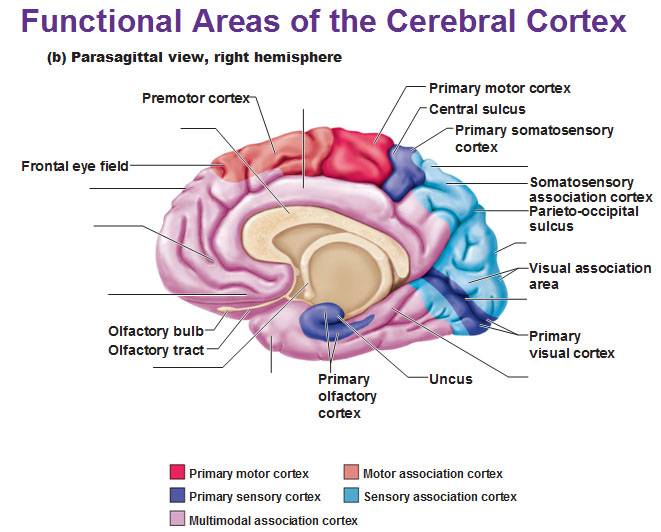 The primary somatosensory cortex sends axons from posterior to anterior. The precentral gyrus is all motor while the postcentral gyrus is all sensory. It’s easy to remember the postcentral gyrus is posterior to the precentral gyrus because it’s in the name: postcentral.

Primary Visual Cortex
Association Areas
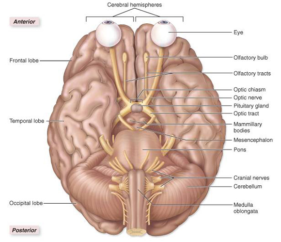
Association areas give meaning to sensations. They are represented by light blue in diagrams. Each primary sensory area has an association area that it projects to. It is what draws upon stored memories to give meaning to sensations such as recognizing the sound of an ambulance siren or a car horn or recognizing the feeling in your pocket are your keys or coins.
Multimodal Association Areas
This is colored pink/lavender in the diagrams. These are large areas of the cerebral cortex that receive sensory input from multiple different sensory modalities and various association areas and help make associations between various kinds of sensory info. We have three multimodal association areas: Posterior, Anterior and Limbic association areas.
The posterior association area is where visual, auditory and somatosensory association areas meet. This is what gives us our spatial awareness of our body. It is the kinesthetic sense that is very strong in professional dancers or athletes for example. On the left side we see the spot in dashes called Wernicke’s area that deals with reading, naming things. On the right side this area helps us to understand emotional overtones/undertones of speech (the multiple ways of saying “I’m fine”).
The anterior association area includes the prefrontal cortex. This part of the brain receives information from posterior association area and helps integrate that info with past experience with the help of the limbic association area. It has a lot to do with thinking and making judgements and it’s where you understand what’s socially acceptable, how to behave; just anything related to heavy human conditioning.
The limbic association area is located on the medial side of the frontal lobe. This is what helps form memories and translates that to motor responses and processes emotions and guides emotional responses. It’s very important for social interactions and expressions of the personality.
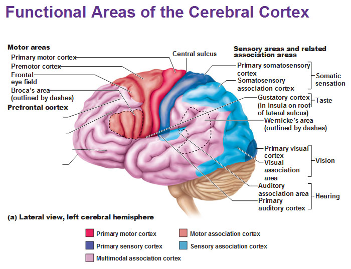
Motor areas, represented by red and dark pink
These include the primary motor cortex, premotor cortex, frontal eye field and broca’s area.
The premotor cortex is located anterior to the primary motor cortex and receives processed sensory information. This helps to plan complex movements and sends the plan to the primary motor cortex.
The primary motor cortex is anterior to the central sulcus and it’s made up of pyramidal cells whose axons runs all the way down the spinal cord to the pyramids in the Medulla Oblongata. Yeah. All the way.
The frontal eye field is anterior to the premotor cortex and helps control voluntary eye movements.
The Broca’s area is the pink outlined area located only in the left cerebral hemisphere. It is anterior to the premotor cortex, near the auditory sensation areas. It is what helps control motor movements for speech production. The corresponding area on the right side controls the emotional overtones to spoken words. In other words, it is what helps dictate how you say/form the words to your speech.
Lateralization of Cortical Functioning
In 90-95% of the population this is the way it goes:
The left hemisphere deals with language, math, science and logic and handles the details.
The right hemisphere deals with visual-spatial skills, reading facial expressions, intuition, emotion, artistic and musical skills and sees the big picture.
The left side sees the trees while the right sees the forest. In the rest of the population this is either reversed or shared equally. Yes, there is a strong correlation between handedness and the hemispherical lateralization as you have always thought.
Use this Table of Contents to go to the next article
YOU ARE HERE AT THE CNS


