Conducting System of the Heart
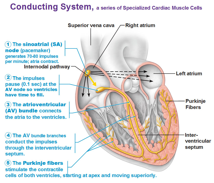
Here’s the deal with cardiac muscle cells. They have this inherent capability (rhythmicity) to contract by themselves unlike skeletal muscle that has to wait for an impulse from a nerve. Cardiac muscle can, if it had to, just contract on its own. It doesn’t necessarily need a nerve to tell it to contract. Look at this picture, this yellow stuff is the actual conducting system which initiates each contraction sequence, which sets the heart beat. These specialized cardiac muscle cells are grouped together in a series that allow them to take on the special job of organizing all that electrical energy. If you didn’t have this system, the muscles would just contract all at once and that’s going to be a mess and we’re going to go nowhere with that. These cells insures that the chambers are going to contract the atria first then the ventricles second, all in a certain direction.
Sinoatrial node (as in sinus, sinus rhythm) is our base rhythm of our heart. This is that 70-80bpm at rest. This SA node sets that rate and so it’s our internal pacemaker. We’re going to send signals to the second node called the atrioventricular (AV) node. We’re going to hold the impulse for a fraction of a section to give the chambers time to fill for a fraction of a section. And then that impulse is going to be allowed to continue and fly down these particular bundles called bundle branches to the apex at the heart (the bottom pointy part) and continue up the sides of the heart at the sides of the ventricles. You see how we have a bunch of terminal cells at the apex? Those are going to be the first ones to fire and then you see the Purkinje fibers which are going to allow blood to contract.

External Innervation
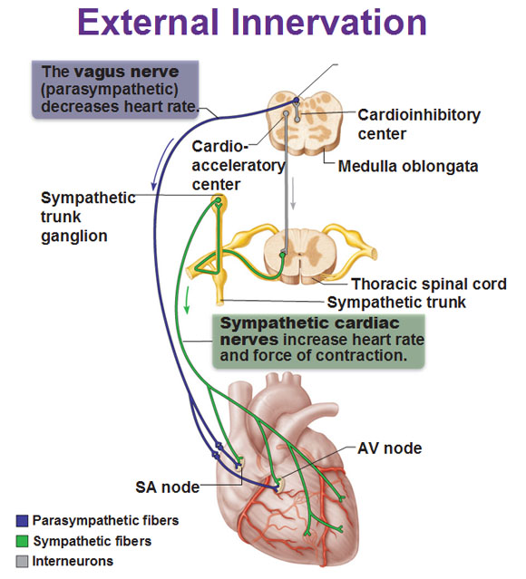
While we have the sinoatrial node providing the base rhythm, the heart rate is altered by external controls. It’s great the SA node can contract at 60-80bpm but if you want to go for a run and these muscles need more oxygen to run, your heart isn’t going to cut it if it’s only going at the same rate to supply all those muscles with oxygen. So we’re going to need an override for the SA node. This is why we even have a nervous system, so that we could do different things and function in real life. The nerves to the heart include the visceral sensory fibers, the parasympathetic branches of the vagus nerve, and the sympathetic fibers from the thoracic spinal cord. The vagus nerve (parasympathetic) coming from the medulla oblongata decreases heart rate while the sympathetic fibers that emanate from the sympathetic trunk increase the rate and force of contraction.
Use this Table of Contents to go to the next article
YOU ARE HERE AT THE CARDIOVASCULAR SYSTEM


