Blood Vessels
Three types of blood vessels that make up the entire system.
Arteries carry blood away from the heart.
Capillaries are the smallest blood vessels where molecules move between blood and interstitial fluid of the tissues.
Veins carry blood toward the heart.
Structure of Blood Vessels
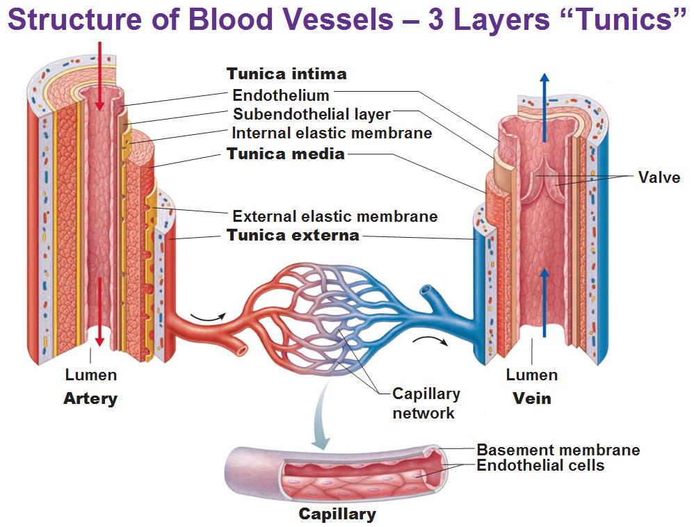
Of course these vessels are not simple tubes so let’s break them down. On your left hand side is a artery, on the right we have a vein. And the capillary on the bottom. We will talk about the artery first.
The inside space where blood flows through is the lumen. The lining of that space is an endothelium made of simple squamous epithelium. Then outside of that is the subendothelial layer. Just outside of that is the internal elastic membrane. If you put these three layers together, it’s a tunic, but specifically it’s called the tunica intima (intima means inside).
Then we have the tunica media (middle layer) which is a pretty thick layer of smooth muscle and elastic fibers. The arrangement of these smooth muscle cells are layers of circles. That tunica media is surrounded by an external elastic membrane.
One more layer is the tunica externa made of connective tissue that’s mostly collagen fibers.
Now let’s look at the veins on the right side. It’s the same layers as the arteries but with different proportions. For veins, they are more-so passively receiving blood. Starting from the inside we have the endothelium made of simple squamous epithelium. We have the subendothelium. We don’t have the internal elastic membrane because they don’t have to stretch and recoil. So we’re going to skip directly to the tunica media which is not as thick. There’s no external elastic membrane either since there’s no internal elastic membrane. Then we have the tunica externa on the outside. And finally, there’s a valve, extending from the endothelium to prevent backflow.
In between them is a capillary bed/network. If you take any part of this network, it’s going to be simple squamous epithelial cells and a very thin basement membrane of epithelial cells.
Some detail on the Tunica Media
The tunica media consists of smooth muscles in the walls of the blood vessels which are directly innervated by the sympathetic division of the autonomic nervous system. When they contract, that’s called vasoconstriction, making the lumen smaller and raising blood pressure. Making the lumen bigger is called vasodilation. There is no direct parasympathetic innervation for vasodilation except for the penis. It’s more of a local response to nitric oxide. Let’s remember, what did we need to happen in the “Flight or Fight” response? What do we need when we need to fight? We need vasodilation at the skeletal muscles to allow more blood to flow. We’ll get back to that, let’s go over arteries.
Types of Arteries
Not all of the arteries are big like the aorta because they keep branching off until they become capillaries. We could separate them in both anatomical and functional terms.
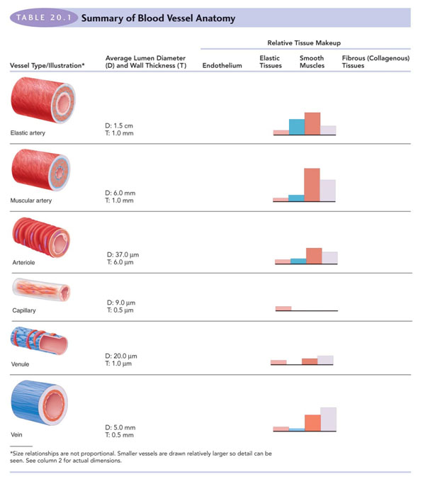
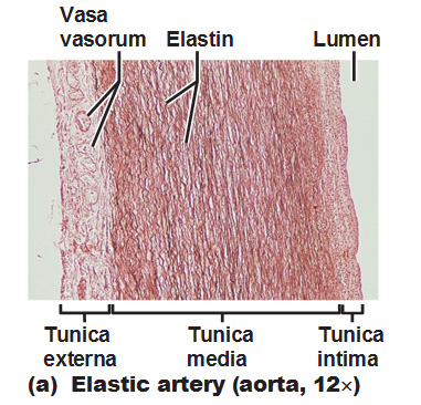 Elastic arteries are the largest, ranging from 2.5cm to 1cm in diameter. They contain a lot of elastic fibers. Showing under the microscope you could see a lot of fibers and it’s a fairly thick layer of tunica media.
The high elastin content in the tunica media layer dampens the surges of blood pressure from heart contractions. The elastin stretches when blood is forced into the artery, stores that energy and during diastole, the fibers recoil, release that energy and propel the blood forward. The blood goes into the next section and that section is going to expand, recoil and help keep it going.
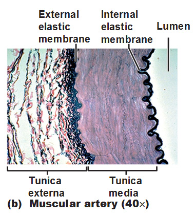 Muscular (distributing) arteries are the medium sized (1cm to 0.3mm) branches that come off the arteries. We’re going to have tunica media but instead of it being full of elastic membrane it’s going to be more smooth muscle and the elastin is concentrated in the internal and external elastic membranes on either side of the tunica media. If we’re going to have a lot of smooth muscle, it means we’re going to have a lot of vasoconstriction and vasodilation. If we have a lot of either happening that means there’s a lot of regulation going on at this particular level. We are controlling a certain amount of blood in that area. Of course we still have elastic fibers (elastin) because we’re dealing with high blood pressure, and large lumens, that will continue to some dampening.
Arterioles are the smallest arteries with diameters ranging from 0.3mm to 10µm. The larger arterioles still contain all 3 tunics but by now that tunica media is only 1-2 layers of smooth muscle cells. Some of them actually, when they start branching, have just one layer of smooth muscle. We’re still doing a little bit of vasoconstriction/dilation, mostly by the local chemicals in tissues and sympathetic nervous system.
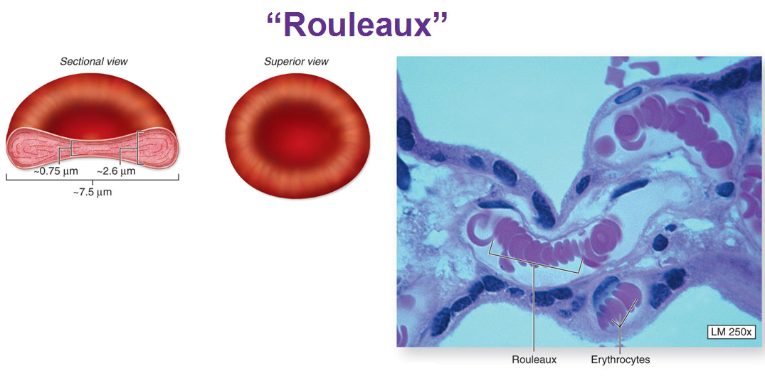
Finally we get to capillaries, these are just barely big enough for a red blood cell to pass through. It’s just a single layer of simple squamous epithelial cells surrounded by a basement membrane. This is where actual delivery of nutrients and oxygen and removal of wastes and carbon dioxide occurs.
Some site-specific functions of capillaries:
Lungs: gas exchange (O2/CO2)
Small intestine: receive digested nutrients
Endocrine glands: pick up hormones.
Kidneys: remove nitrogenous wastes.
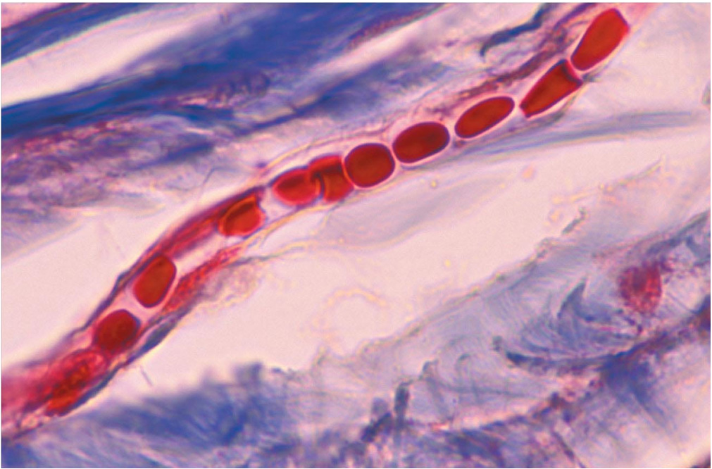
Red Blood Cells in a capillary:

This is a Rouleaux (roll of or stacked up coins) that occurs when there is a problem:
Capillary Bed
There’s a terminal arteriole (that sends blood to the capillaries) and a postcapillary venule (that receives blood from the capillaries). If you really look at it, there’s a central passage way and there’s all these little capillaries coming off of it but there’s definitely a main street between the arteriole and venule. The beginning of this main streem is called a metarteriole. The entrance to side streets (true capillaries) have these little collections of smooth muscles and function as precapillary sphincters which open and close. The distal portion of a metarteriole has no smooth muscle sphincters and is called a thoroughfare channel. In this particular picture you could see the left side has a lot of oxygen while the right side is diffusing Carbon dioxide out the right side.
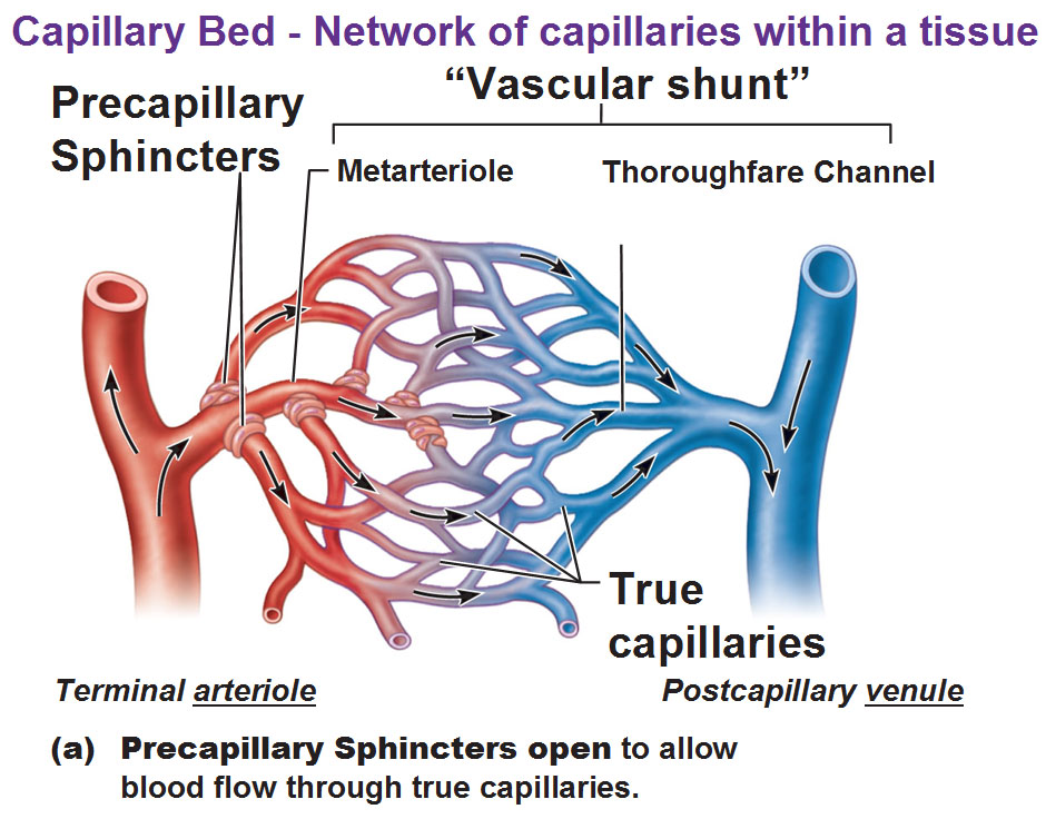
Let’s compare that with this next picture. The sphincters have contracted to close off the side streets so the blood goes through the thoroughfare channel and head over to the venule. You could still have gas or nutrient exchange but it’s going to pass a lot faster because the blood isn’t meandering and traveling all over the place. The blood is just going to be shunted through that area. When would you have this scenario so the blood would get shunted away from certain organs into another? During the sympathetic (flight or fight) system. When you’re flighting/fighting it shuts blood away from the digestive system to divert resources to the skeletal muscles.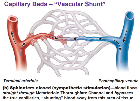
One last note: Remember there were certain tissues that didn’t have very many capillaries or none at all in them, remember? Things like tendons, ligaments, cartilage, cornea, lens and epithelia are poorly vascularized and depend on the tissues that are next to them and wait for them to diffuse oxygen and nutrients over to them.
Different structures of capillaries determine the degree of permeability
Function and structure are very intimately tied in together.
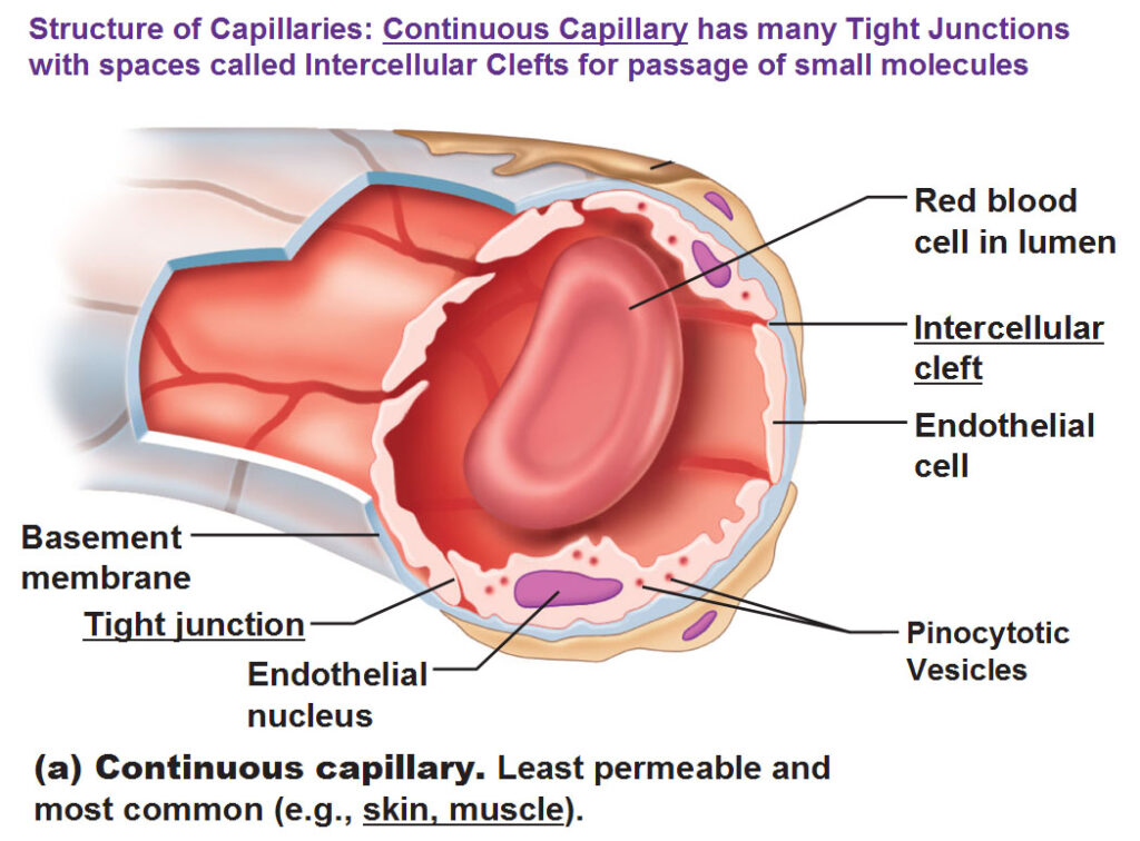
This is called a Continuous Capillary. The spaces in between the individual endothelial cells are loaded with tight junctions holding the cells together of a continuous type of capillary. The spaces in between that allow things to pass through are called intercellular clefts that are not totally sealed but very tiny making these the least permeable capillaries and most common. This is found in skin and muscle, sort of like a standard issue capillary.
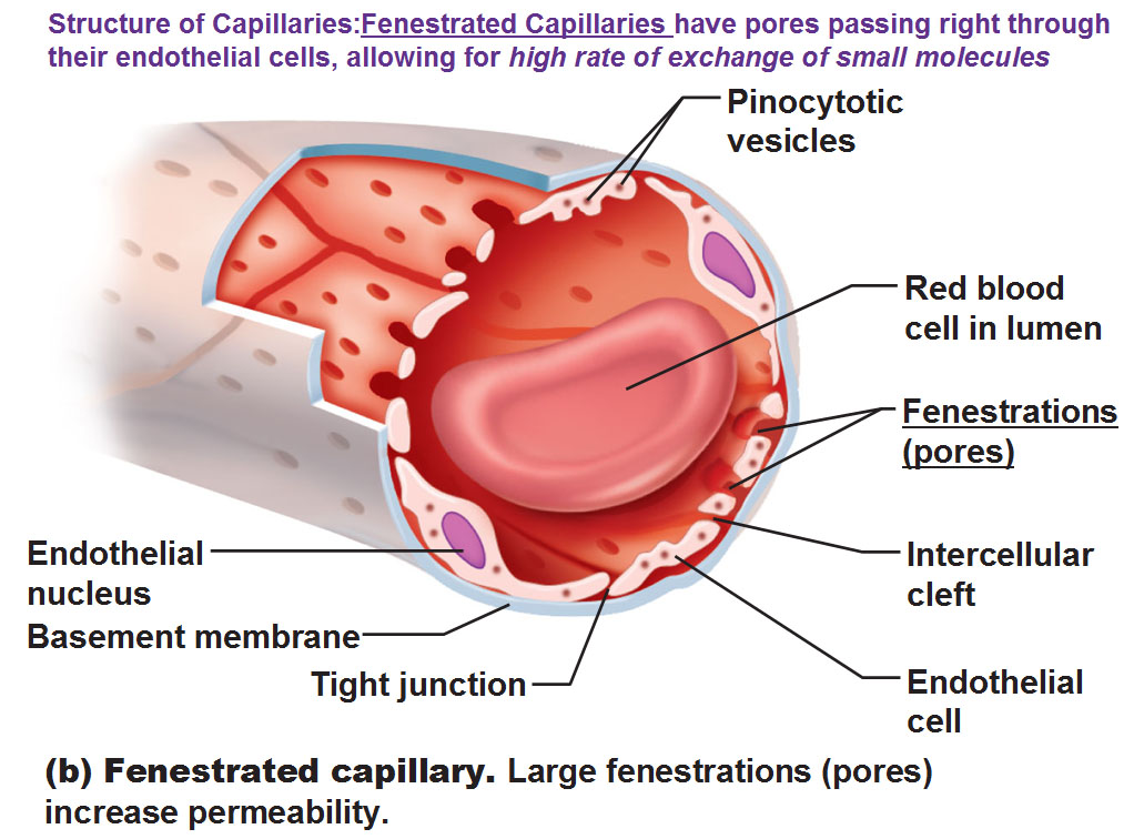
Next we have Fenestrated capillaries. Fenestrated just means holes, they literally have holes that pierce these endothelial cells so we could have small molecules pass through easily. We still have tight junctions but not as many so the spaces between cells are going to be a little bigger. This is specifically found in the kidneys (lots of filtration of fluid so you want stuff to pass) and small intestine (digesting lots of stuff and nutrients need to flow quickly) because of what they need to do.
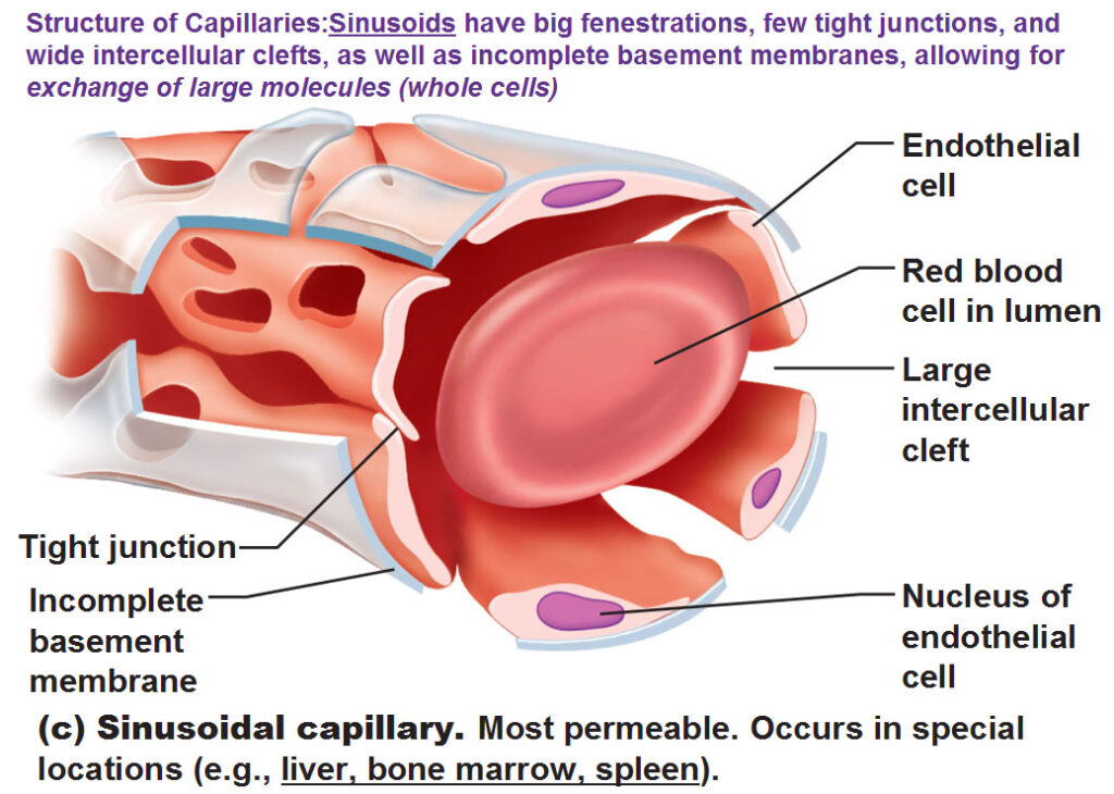
Next is a Sinusoid that allows entire cells to pass through. This capillary has big fenestrations with hardly any tight junctions so we have large intercellular clefts in between those endothelial cells. Also on top of it we have missing sections in the basement membrane so it’s an incredibly very loose structure and it’s found in the bone marrow, liver and spleen because you want to have a lot of free flow. The liver is for detox. Bone marrow is for the blood cells. The spleen has cleaning up duties (viruses, bacteria, toxins, old blood cells, etc).
The capillaries in the Blood Brain Barrier have tight junctions around the entire perimeter of endothelial cells so that there are no intercellular clefts. Only the most vital molecules pas through via highly selective cell membrane transport mechanisms. However, it’s not a barrier against lipid-soluble molecules such as oxygen, co2, anesthetics and other drugs. During extreme and prolonged states of stress, the tight junctions will come apart and open allowing toxins to cross into brain tissue.
Veins
Veins conduct blood from capillaries toward the heart. Our smallest veins are venules which join together to get bigger and bigger to eventually lead into the vena cava. Our smallest venules are the postcapillary venules. The following picture is just a comparison of the different levels of arteries and capillaries and veins and different thicknesses of tunics as you go from one structure to another and how those tunics shift in thickness. Notice how the elastic and muscular arteries have a lot of smooth muscle and then look at the bottom at the vein. The tunica externa is the thickest tunic in the veins.
Mechanisms to counteract low venous pressure and help blood go back to heart
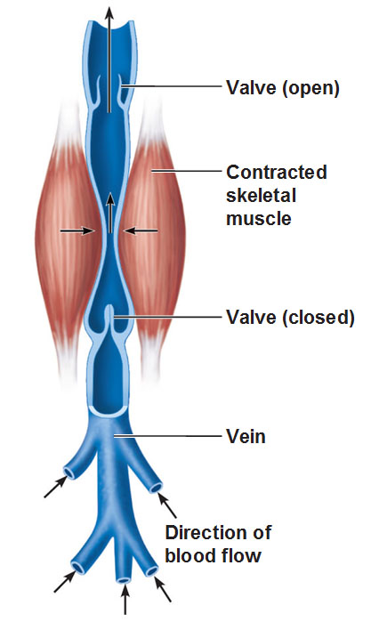 Well, by the time you get to the veins, the pressure from the heart beating has dissipated. We have to practically get this blood back to the heart. So we have these valves layered on the inside while on the outside we have skeletal muscles and as we move, our muscles squeeze and massage the veins and work the blood back up to prevent it from pooling down at your feet. This has a name of course: the skeletal muscle pump and the valves are arranged in such a way that as one bolus of blood goes through if it starts to fall, it shuts down to prevent backflow. When these valves get overworked or strained, they stretch out the vein wall permanently. A classic example would be… when a woman is pregnant, it’s harder for these valves to hold the pressure in the pelvis and blood may pool in a section and if it doesn’t recoil, that section of bulge becomes varicose veins.
Vasa Vasorum is the blood supply of the larger blood vessels themselves, such as the aorta. These large vessels have tiny arteries, capillaries and veins in their tunica externa. They come from either the same vessel or a nearby vessel and nourish the outer half of the vessel wall. The inner half receives nutrients from luminal blood.
Vascular anastomoses is a concept where an area will receive blood from two completely different arteriole (or venous) branches so if one branch is cut off or injured that one particular area will still receive nourishment or oxygen from the other branch. Most organs receive blood from more than 1 arterial branch and when neighboring branches connect with each other, that’s anastomosis. We have lots of anastomoses around joints, abdominal organs, brain and heart. Veins anastomose even more than arteries.
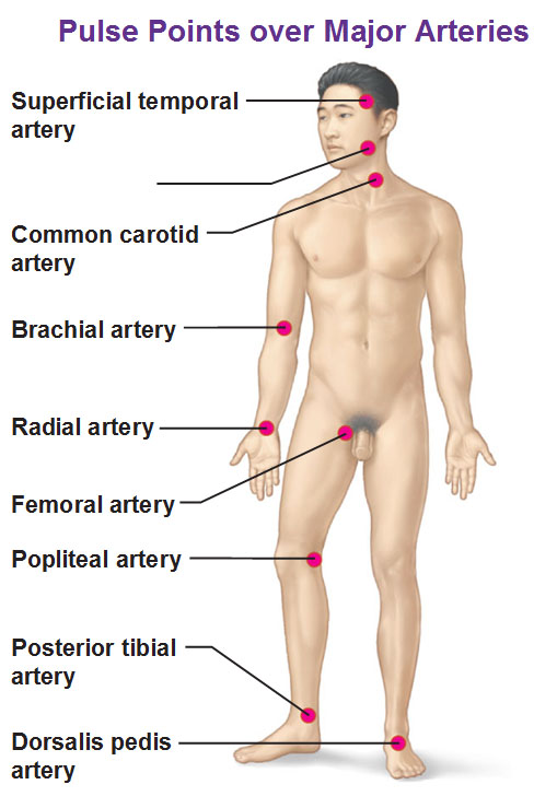
Pulse points over major arteries
The most common are the common carotid and radial artery.
Femoral artery is the easiest to feel as it’s very easy to feel because it’s big and close to the surface.
The posterior tibial artery and dorsalis pedis artery are very important for very old people to check if they are getting blood through there.
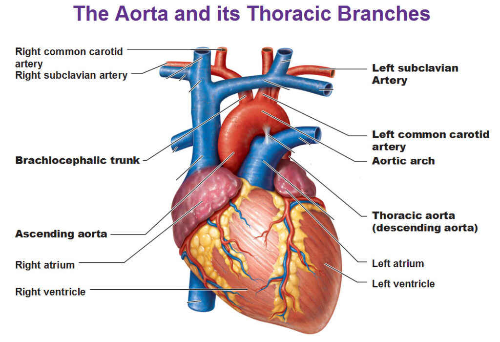
The Aorta
The ascending aorta is the part that goes upward directly out of the heart. The descending aorta is the one tucking down behind the heart.
The first branch right at the top of the aortic arch is the brachiocephalic trunk and that’s going to go off to the right side of the body and quickly branch to the right common carotid and right subclavian.
The second branch off the aortic arch is the left common carotid artery. (There is no left brachiocephalic trunk). On the left side we don’t have a brachiocephalic because it’s already closer to the left side and it’s close enough to the arm so you don’t need any brachiocephalic business but on the right side you need it before it branches off.
The third branch is the left subclavian artery.
Major arteries of the head and neck
Below is a lateral view on the right side of our head. Here’s our brachiocephalic trunk from a different view and it goes to the subclavian and the common carotid up towards the head. The thing that’s unique in this particular drawing is the vertebral artery running through the transverse processes of the cervical vertebrae.
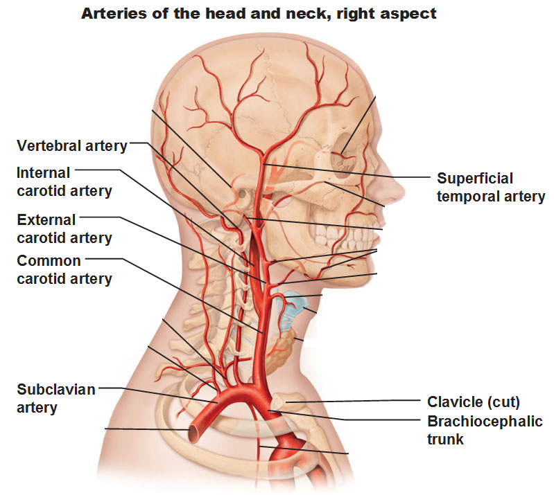
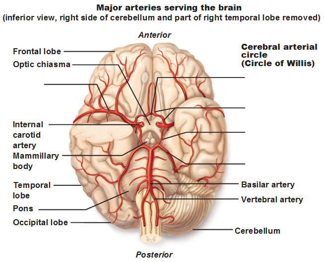
Below: Look at the vertebral artery making its way inside, up along the brainstem (the medulla oblongota specifically) and it’s going to come together at the Pons and be called the basilar artery when it joins.
Use pictures Above and Below: Meanwhile, coming from common carotid, if we follow it from the image above, it branches into the external carotid and internal carotid. The internal carotid comes into the brain from underneath the temporal lobe. The basilar artery is going to send branches to the carotid artery and vice versa and we get a nice circle called the Circle of Willis and this is the vasa anastamoses that we talked about earlier. We have stuff coming from the vertebral and carotid arteries together to literally create a circle so if one of those arteries get cut or blocked there’s still other stuff that supplies this area. This idea of anastomosis is going to be seen over and over in our vital organs.
Arteries of the Upper limb and Thorax
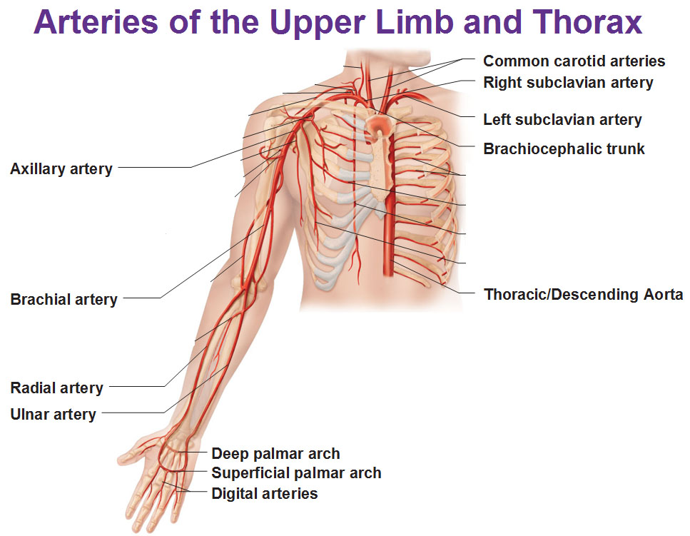
We see our major branches off the right subclavian such as the axillary, brachial and radial and ulnar arteries. This radial artery is one of the major pulse points.
EVERYTHING FROM THIS POINT ON IS UNREVISED AND UNFINISHED DUE TO LACK OF TIME.
Renal artery named for where it’s going to (renal always has to do with the kidneys).
The celiac trunk is a branch off the abdominal aorta and there’s the left and right gastric artery that goes to the stomach (Gastric=stomach). We also have the left and right gastroepiploic artery that is this great curvature along the stomach supplying blood there. Let’s keep going..
Superior Mesenteric Artery
Mesentery holds the small intestine in place and the mesentery helps distribute this particular artery to the small intestine. Here is them branching off the aorta from the common iliacs. (slide 3 part 3) is a detail of this. External iliac continues… (to be continued).
Venous system
a lot of them are running along the arteries and often have the same names as the arteries which we are all grateful for.
Veins of the head an neck: Here’s our drainage of the head and neck . For example we have the vertebral vein that runs along side the vertebral artery. Instead of carotid we have jugular veins. Superior dumps into the right atrium
When we are talking about veins we are talking about tributaries. When we are talking about arteries they are called branches. Your inferior vena cava leads up to your right atrium and that’s receiving stuff from everything below (your heart) and that’s coming from your hepatic veins (coming from where?????????) and the renal veins coming from the kidneys.
Basic Scheme of the Hepatic Portal System is a very specific, special part of the venous system that’s specifically all of these veins that drain the small intestine but here specifically take the nutrients and deliver them directly to the liver. Hepatic (liver) Portal (like a doorway) system. And we want to take all that stuff from the intestines and into the liver first because all that stuff came from the outside that we want the liver to clean up first before we deliver to the heart and infect the heart or distribute it to the rest of the body. So we want to clean up the blood and then go toward the heart. The hepatic portal vein takes the stuff, brings it to the liver and the hepatic veins are the ones that drain the liver and deliver the processed blood to the inferior vana cava.
Use this Table of Contents to go to the next article
YOU ARE HERE AT THE CARDIOVASCULAR SYSTEM


