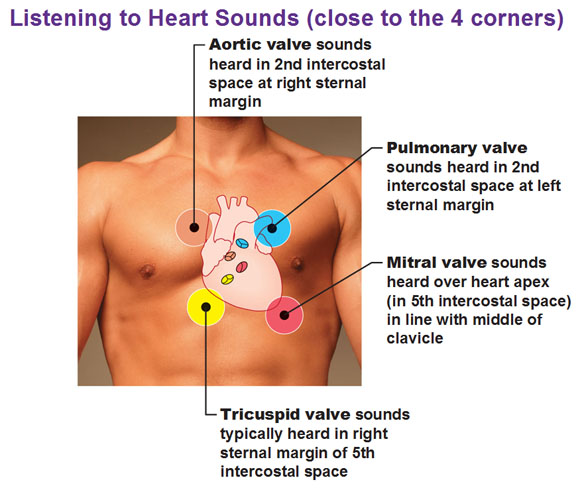
The four corners of the heart

When we look at the surface of the heart, we could sort of imagine it like a square with four “corners.” Why would we even try to define the four corners of the heart? Because each of those corners correspond to the best spot where you could put your stethoscope and hear each valve better than the other three. So if you put your stethoscope right over the mitral corner, you’re going to hear the mitral valve stronger than all the other valves. You’re still going to hear the other sounds, but you’ll focus your attention onto that mitral valve because that’s going to be the clearest at this location. The same goes for all the other locations. That’s why you want to learn the four corners of the heart.
 We have two on the right, two on the left, divided into superior and inferior labels.
Superior right: At costal cartilage (cartilage where ribs attach to sternum) of the right third rib and the sternum.
Inferior right: At costal cartilage of right sixth rib, one finger’s width lateral to the sternum.
Superior left: At costal cartilage of left second rib, one finger’s width lateral to the sternum.
Inferior left: Lies in the fifth intercostal space (below 5th rib) at the midclavicular line.
Use this Table of Contents to go to the next article

YOU ARE HERE AT THE CARDIOVASCULAR SYSTEM


