Review of the Nervous System

In any physiology book the nervous system takes up a third of the book and it’s the most complicated because it controls everything else. Let’s review the basic anatomy of the nerves before we jump into the heavy topics. If any of this is new to you, then please review the Anatomy posts about the Nervous System.
Central Nervous System and Peripheral Nervous System
We divide the nervous system into two parts, the central nervous system and the peripheral nervous system. The central nervous system (CNS) is the brain and spinal cord and these are the most protected parts of your body. The brain is enclosed in the skull and the spinal cord is enclosed in the vertebral column.
The peripheral nervous system (PNS) refers to all the nerves branching off of the brain and spinal cord. We have 31 pairs of spinal nerves coming off the vertebrae and 12 pairs of cranial nerves coming off the brainstem.
The 31 pairs of spinal nerves consist of…
- 8 pairs of cervical
- 12 pairs of thoracic
- 5 pairs of lumbar
- 5 pairs of sacral
- 1 pair of coccygeal nerves
The identification of the spinal nerves are based on the location in which they exit from the spinal column. These are all pretty easy to remember because they all respond to the number of vertebrae. While there is only one sacrum, it’s actually 5 bones fused together which is why there are 5 pairs of sacral spinal nerves.
As for the discrepancy of having 8 pairs of cervical spinal nerves and only 7 cervical vertebrae? The first pair of spinal nerves comes off from between C1 and the Atlas, so you end up with a total of 8, even though we have 7 cervical vertebrae.
When we talk about a nerve, think of it like a telephone cable. A telephone cable actually has several wires in it. Inside each nerve are hundreds of thousands of very thin nerve cells (interchangeably called nerve fibers because they are long just like muscle fibers.)
Each nerve may include sensory (afferent) nerve fibers and motor (efferent) nerve fibers.
- The sensory fibers relay info from the organs to the CNS.
- The motor fibers relay orders from the CNS to the organs.
The Meninges
The meninges are membranes that ensheath the CNS to protect it. The outer one is dura mater (tough mother) made of dense regular fibrous connective tissue that provides a tough barrier against foreign agents. The middle membrane is the arachnoid membrane. The one below it is the pia mater (soft mother) that adheres to the surface of the brain and spinal cord. Between the arachnoid and pia mater is the subarachnoid space that is filled out with CSF. Just outside the dura mater is the epidural space. If you’ve ever heard of a woman having an epidural (lumbar puncture), a woman has it inserted just outside that membrane to tap into the CSF.
Ever heard of spinal meningitis? This is a debilitating condition that involves the inflammation (-itis) of the meninges that spreads. When damage occurs, it is irreversible/irreplaceable.
Dermatomes
A dermatome is an area of skin innervated by the cutaneous branches of a single spinal nerve. If someones been in an injury and they can’t feel that area, then it’s likely that nerve is affected. The cervical spinal nerves primarily innervate the neck and the arms, the thoracic spinal nerves innervate the chest and torso, and the lumbar spinal nerves primarily innervate the legs. Look at my dermatomes post to see a map of which nerves innervate which part of the body. parts of the body are innervated by the nerves. If you look a the thumb it’s labeled C6 for example. You have a right and left thumb but you also have a pair of these C6 spinal nerves.
Supporting cells of the CNS (Glial Cells)
Relevant Anatomy Post: Review the Fundamentals of the Nervous System and Nervous Tissue
Not all the cells of the nervous system are nerves. So-called glial cells of the CNS account for over half the cells in your brain. Textbooks usually identify 6 different types of glial cells. We’re going to review 3 of them.
1. Oligodendrocytes: They form a myelin sheath around nerve fibers. Analogy: You know how you could have a bare copper wire or a copper wire with a plastic coating? We have nerve cells that are bare and also nerve cells that have the equivalent of a plastic coating. Instead of a plastic coating it’s a myelin sheath. The lipoprotein in a myelin sheath is called.. myelin which helps insulate the axon to prevent interference from nearby neurons and increases the impulse speed.
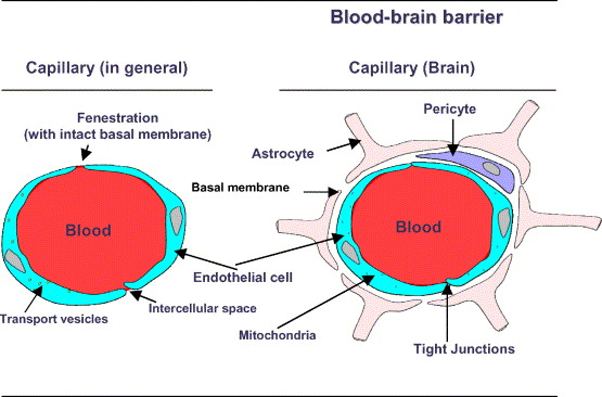
2. Astrocytes are cells that surround the capillaries of the CNS and create a barrier called the Blood-Brain-Barrier. Look at the picture below where we have two capillaries. The one on the left is a general typical capillary found anywhere in the body. The wall of a capillary is made up of a single layer of cells (simple squamous epithelium) and blood flows through these delicate vessels. All the chemicals can easily diffuse across the capillary walls. In the capillaries of the CNS, there’s an additional layer of cells that wrap themselves around the outer surface of the capillary and these are called astrocytes. The capillaries of the CNS are essentially two-cells-layer thick which creates an additional barrier between the neurons and what’s in the blood stream as an added layer of protection.

3. Microglia are small phagocytes that swallow up bacteria and viruses that are in the CNS. When bad guys invade your body, white blood cells get involved.
When somebody is robbing your home, you call the police and they arrive in an hour and everyone’s dead and they take a report for two reports. Who does the president rely on? The secret service. The secret service has an oathe to jump in front of a bullet to protect the president. Like the president, the brain has its own secret service squadron.
What should impress you are the layers of protection: We have the skull. The 3 layers of membranes (dura, arachnoid, pia). All these additional astrocytes. Some of these nerves have these myelin sheaths. It’s got its own squadron to protect from bacteria and viruses.
Supporting cells of the PNS (Schwann Cells)
What are schwann cells? Schwann cells are to the PNS what the oligodenroglia are to the CNS. Schwann cells form the myelin sheath of the sensory or motor neurons in the peripheral nervous system. Oligodendrocytes wrap around and form the myelin sheath for all the nerves in the CNS.
Now let’s review neurons…


