Microbiology Tests

Mannitol Salt Agar
Mannitol provides the substrate for fermentation and makes the medium differential. The salt makes the medium selective because its concentration is high enough to dehydrate and kill most bacteria. Staphylococci thrive in the medium largely because of their adaptation to salty habitants, such as human skin. Most Staphylococci are able to grow on MSA but don’t ferment the mannitol so the growth appears with the unchanged pink or red medium color. S. aureus, however, ferments mannitol, which produces acid and lowers the pH of the medium. The result is bright yellow colonies. This test is specifically for isolating and differentiating S. Aureus.
- If there is no growth at all, the salt inhibited the organism, it’s not Staphylococcus.
- If there is good growth, the organism is not inhibited by the salt and it’s Staphylococcus.
- If there is good growth AND THERE’S A YELLOW HALO AROUND IT, then this organism is not inhibited AND it ferments mannitol, so it’s possibly S. Aureus!
- Moral of the story: If you see yellow, it’s probably S. Aureus.
Bile Esculin Test
In this medium there is bile and esculin. Bile is the selective agent used to separate Streptococcus bovis and enterococci from other streptococci. Esculin is present because only the group D streptococci and enterococci can hydrolyze esculin in the presence of bile salts.
- Any blackening of the medium means Bile-Esculin positive and presumptive identification of group D streptococcus or Enterococcus.
- Note, if there is blackening of the medium before 48 hours, it can be recorded as Bile-Esculin positive.
- If there is no darkening after 48 hours, it is not a group D streptococcus or Enterococcus and is Bile-Esculin negative.
- Incubation for at least 48 hours must pass for the negative result to be determined.
MacConkey Agar
This is a selective and differential medium for the growth of gram-negative bacteria and contains lactose and it’s dark brown colored. If the bacteria on it can ferment the lactose, the acidity turns the dye into a bright pink color.
- If there is poor or no growth, the organism is inhibited and it’s probably gram-positive.
- If there is good growth, then it’s gram-negative.
- If there is good growth AND it’s red, this means the gram-negative bacteria is producing acid from lactose fermentation and it’s probably a coliform like E. coli.
- If there is good growth but it’s not red, then it’s a noncoliform bacteria.
Eosin Methylene Blue Agar (EMB Agar)
This is a complex, selective and differential medium used to isolate fecal coliforms. The dyes inhibit the growth of gram-positive organisms (like the MacConkey) but they also react with lactose fermenters, turning the growth dark purple or black. If it’s a black growth with green metallic sheen, it’s likely E. coli. If it’s a pink color, it’s a less vigorous lactose fermenter. If there is a lack of color, it’s a nonfermenter.
- If there is poor or no growth, the organism is inhibited and it’s probably gram-positive (like the MacConkey)
- If there is good growth, then it’s gram-negative (like the MacConkey)
- If there is good growth and it’s “dark” purple or black, it’s probably a coliform like E. coli.
- If there is good growth AND it’s red/pink, it’s probably a coliform like Enterobacter or Klebsiella.
- If there is good growth but it’s not pink, purple or metallic and there’s no color, it’s a noncoliform bacteria (because it doesn’t ferment lactose or sucrose).
Thioglycollate Medium
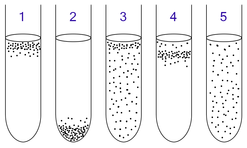
Normally this broth looks clear/straw-colored and the top is slightly pink due to oxidation of the dye with its contact to oxygen. This medium can be used to grow microbes representing all levels of OXYGEN TOLERANCE but it’s generally associated with cultivating strict anaerobes and microaerophiles. A microaerophile requires oxygen to survive, but environments containing lower levels of oxygen than the atmosphere.
So let’s go over what these results mean:

- If only the top has growth, that means it is a strict aerobe because it is growing only in the top part.
- If the bottom has growth, it means it’s a strict anaerobe (aka obligate anaerobe) because it can grow in the portion furthest away from the oxygen.
- If there is growth throughout, but mostly at the top, it is a facultative bacteria since it likes aerobic respiration best but can grow without it as well.
- If there is only growth just below the very top, that’s indicative of a microaerophile, because it needs oxygen to live, but not a lot of oxygen, so it’s not going to grow closest to the top.
- If there is growth throughout evenly diffused, that’s indicative of an aerotolerant anaerobe as the presence of oxygen doesn’t affect them.
Oxidation-Fermentation Test (O-F)
This test is designed to differentiate bacteria on the basis of fermentative or oxidative metabolism of carbohydrates. In fermentative pathways, when the carbohydrate is broken up, there is acid production present. Two tubes are used, one with mineral oil to promote anaerobic growth/fermentation and the other has no mineral oil to allow aerobic growth and oxidation.
- The control is green. If both tubes turn yellow, it means the organism can ferment the carb AND oxidize the carb in both scenarios.
- The control is green. If both tubes remain green, there is no sugar metabolism and the organism is nonsaccharolytic.
- If the sealed tube remains green, but the unsealed becomes yellow, it has oxidative fermentation.
- If there is slight yellowing only at the top of both tubes, it’s oxidative and/or slow fermentation.
- Note: A fermentative organism (F) will look exactly the same as an organism capable of both oxidation and fermentation (O-F).
- Oxidation = Pseudomonas and Alcaligenes
- Fermentation = E. Coli (facultative)
So it’s very easy, in a O-F tube, it starts as green, any yellowing indicates the breakdown of carbs and whether it has mineral oil or not determines under what conditions the bacteria can do it under.
Phenol Red Broth
This is a broth to which a carb such as lactose or sucrose is added and there is a durham tube that is used to indicate gas production.
Methyl Red and Voges Proskauer Tests
The MR test is used to detect organisms capable of performing a mixed acid fermentation.
MR and VP are the same media to start with.
- MR: E. Coli is positive. E. aerogenes is negative.
- VP: E. coli is negative. E. aerogenes is positive.
Catalase
- Catalase positive = Staphylococcus epidermidis
- Catalase negative = Enterococcus faecalis
Nitrate Reduction
- Gas presence; nitrate positive = E. coli
Citrate Test
Used to determine if the organism has the ability to use citrate as its sole source of carbon. Part of the IMViC (Indole, Methyl Red, VP and Citrate) tests to differentiate members of Enterobacteriacea and other gram-negative rods.
- Positive if blue = Enterobacter aerogenes
- Negative if green = E. coli
Decarboxylation Test
Just like the citrate test, it’s used to differentiate organisms in the Enterobacteriaceae family.
- Purple is the only positive result and indicates that the organism produces the decarboxylase enzyme.
- Enterobacter aerogenes is positive.
- All other results are negative.
Phenylalanine Deaminase Test
Organisms that produce phenylalanine deaminase can remove the amine group (NH2) from the amino acid phenylalanine. The phenylalanine agar is used to differentiate the genera Morganella, Proteus and Providencia from other members of the Enterobacteriaceae.
The agar is normally yellow. If the phenylalanine deaminase is present, then when phenylalanine is broken down, there will be phenylpyruvic acid present and when you add ferric chloride, it will turn green.
- A dull military green color is a positive result indicating the presence of phenylalanine deaminase.
- Proteus vulgaris is PD positive.
- No color change means PD is absent.
- E. coli is PD negative.
Urea hydrolysis
Urea is a product of decarboxylation of certain amino acids. It can be hydrolyzed to ammonia and CO2 by bacteria containing the urease enzyme. Urea agar was formulated to differentiate rapid urease-positive bacteria from slower urease-positive bacteria and urease-negative bacteria.
Control looks orange.
- If it turns ALL pink within 24 hours, that indicates rapid urea hydrolysis; strong urease production.
- If it turns partially pink within 24 hours (or all pink after 24hrs), that indicates slow urea hydrolysis.
- If it remains orange within 24 hours and then all or partially pink after 24hrs, that is also slow urea hydrolysis.
- If it remains orange or yellow, no urea hydrolysis; urease is absent.
- E. coli is urease negative.
- Proteus vulgaris and Klebsiella pneumoniae are urease positive.
Gelatin Hydrolysis Test
This test is used to determine the ability of a microbe to produce gelatinases. If the gelatin turns to liquid, gelatinase is present.
- S. Aureus is gelatinase-positive and can be differentiated from S. epidermidis.
- Serratia and Proteus are gelatinase-positive and can be differentiated from other enterobacteriaceae.
- Bacillus anthracis, B. cereus are gelatinase positive, as are clostridium tetani and c. perfringens.
- E. coli is gelatinase negative.
Triple Sugar Iron Agar / Kligler Iron Agar
You have to know how to read slants and butts. You typically read the slant first, then the butt.
- A yellow color indicates fermentation with acid accumulation
- A pink/red color indicates protein catabolism with alkaline products.
- Black precipitate indicates Hydrogen Sulfide (H2S).
- A/A+ = Acid (yellow) slant / Acid (yellow) butt
- K/A = Alkaline (red) slant / Acid (yellow) butt
- K/K = Alkaline (red) slant / Alkaline (red) butt = Not enterobacteriaceae
- K/NC = Alkaline (red) slant / No change in butt = Not enterobacteriaceae
- NC/NC = Organism isn’t growing at all = Not enterobacteriaceae
- H2S = Black precipitate indicates sulfur reduction.
Summary of what we know about E. coli
- E. coli grows pink/red in MacConkey agar
- Grows dark purple with a green metallic sheen on the EMB agar,
- it’s fermentative (facultative) in the O-F test,
- positive in the Methyl Red test,
- negative in the VP test,
- nitrate positive,
- citrate negative,
- phenylalanine deaminase negative,
- urease negative,
- gelatinase negative


